
LIV.INNO students apply their skills to Adaptix innovation project
40 per cent of scaphoid bone fractures in the wrist are missed in a 2D x-ray, sometimes resulting in permanent damage for the sufferer and litigation for the hospital.
Now a new 3D x-ray technology from Adaptix has potential to cost-effectively provide greater visibility for a more accurate diagnosis. Using powerful computing and simulations, the company is looking to extend its imaging to chest x-rays with the help of the Liverpool Centre for Doctoral Training for Innovation in Data Intensive Science (LIV.INNO).
Professor Carsten P Welsch, Director of LIV.INNO, has just been awarded £400k from STFC to design a new system for chest imaging. The project will use AI and Machine Learning techniques, developed by students and staff from LIV.INNO.
Dr Steve Wells, chief technical officer at Adaptix, explained: “2D x-rays are a bit like a shadow puppet – everything is superimposed, so it is difficult to understand in detail what is really going on. CT (computed tomography) and MRI (magnetic resonance imaging) provide much better images, but they are expensive and, in the case of CT, also delivers a much higher dose of radiation.
“The challenge is how to provide cost-effective, high resolution 3D images with a mobile scanner that delivers a low dose of radiation,” Dr Wells continued. “We have been working with the QUASAR Group at University of Liverpool to develop scanners that fire x-rays from an array of positions to do this.”
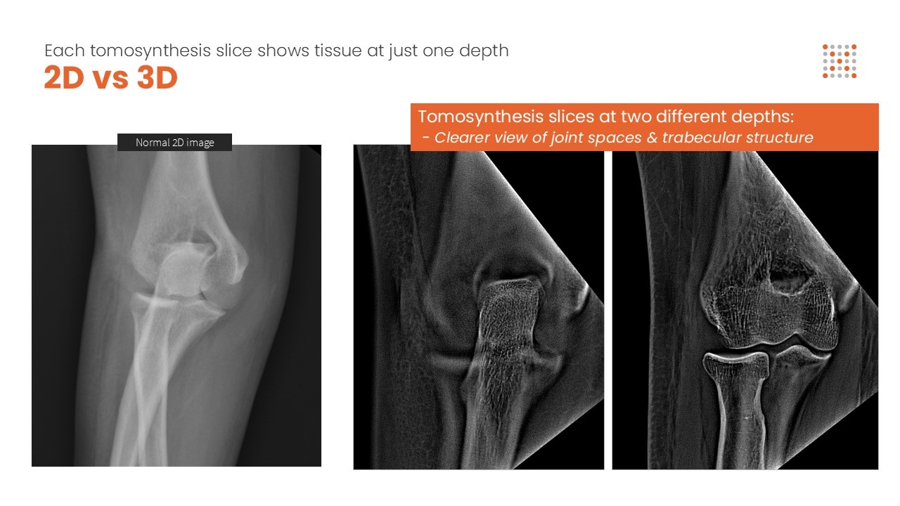
The Adaptix technology uses digital tomosynthesis, where x-rays are fired sequentially from many angles in order to create slices through the object at different depths. From this, anatomy in 3D can be visualized, with an order of magnitude less radiation than a conventional CT -scan.
The potential of the technology has already been recognised by the NHS. Dr Wells’ team has recently been to the Isles of Scilly to investigate the potential of a mobile scanner improving access to 3D imaging for remote communities.
Dr Wells continued: “Scaphoid injuries are commonly caused when you try and save yourself from a fall with an open hand. If someone falls on one of the little islands they need to take a boat to the main island – and the radiographer is only there on a Thursday! A scanner that could be used by a specially-trained paramedic would save the cost of flying someone to the mainland and enable much more rapid treatment.”
The technology is producing images that have been well received by radiologists and the company is working on regulatory approvals.
Dr Wells has been working with the team headed up by Professor Carsten P Welsch at the University of Liverpool for over a decade to refine the imaging technology.
More recently Professor Welsch has established LIV.INNO to offer students comprehensive training in data-intensive science to help develop their knowledge of AI, machine learning, data analysis and the Monte Carlo method for modelling complex radiation fields. Within the training they get the opportunity to apply their skills to real-world challenges such as those faced by Adaptix.
Professor Welsch explained: “There is a shortage of skills in AI; our demanding industry projects enable the co-development of solutions to intractable problems, creating competitive advantage for the companies and invaluable commercial skills and experience for our staff and students.”
Dr Wells agreed: “There are a huge number of variables when designing emitter array systems and a lot of interdependencies. It is good to collaborate with data science experts from Liverpool who are just itching to deal with the sort of headaches we have encountered.
“Initially we did not get very far in our own brute force attempts of multi-parameter system optimisation.
“The Liverpool team applied a much smarter approach by modelling the system using genetic algorithms, where each parameter is considered a gene and a combination of parameters a chromosome. Sending these off to the computing equivalent of ‘Love Island’ enabled us to narrow down the most optimal solutions more quickly.”
Use of simulation also lowers the cost of physical prototypes as the images are of sufficient quality to show to radiologists before committing to hardware development.
The scale-up of the technology to enable improved scanning of the lungs involves adding further elements of complexity, but a computer modelling approach will save a significant investment in hardware.
The innovation of bedside 3D chest imaging offers many exciting prospects, such as increasing confidence in the placement of naso-gastric tubes, detection of pneumothorax, and regular, low-dose monitoring of conditions such as cystic fibrosis. It could also help with early detection of lung cancer.
Dr Wells continued: “Traditional 2D chest x-rays are not good at spotting lung cancer in its early stages when it could be treated more effectively. Therefore, the disease is often not being diagnosed before it has spread, so today for every eight people diagnosed, on average only one will be alive in five years’ time.
“Our technology has the potential to provide early diagnosis with a mobile 3D x-ray scanner, making a quality diagnosis more widely accessible.”
Adaptix have already sold systems for imaging small animals in veterinary practices and also for non-destructive testing (NDT) of electronics and composites. Adaptix will be showing its NDT technology at the Advanced Engineering Show at the NEC Birmingham on 30-31 October 2024.