
European researchers are developing a new real-time scanner that will create a full image of a moving eye without blurring. Teaming up with photonics innovation hub ACTPHAST 4R, the scientists at Vrije Universiteit (VU) Amsterdam are progressing their scanner concept to a demonstrator stage to acquire data faster than existing optical imaging technologies.
Some degenerative eye conditions like glaucoma, diabetic retinopathy, macular holes and retinal diseases can progress to blindness if they are not diagnosed in their early stages due to missed opportunities from poor image quality or motion artefacts. Eye specialists currently use an imaging technique called Optical Coherence Tomography (OCT), a non-invasive test that uses light to build up an image of the retina by capturing cross-sectional slices. However, because the eye is constantly moving, the images suffer from blurring and often only partial pictures are possible. OCT technology has never been fast enough to take a full image of a moving eye without blurring or expecting the patient to sit incredibly still.
The lead researcher on this breakthrough development, Assistant Professor Imran Avci from the Department of Physics and Astronomy at VU Amsterdam, said: “Diagnoses of eye diseases that could lead to blindness require good quality images at an early stage. Our scanner is different: with the data acquired fast enough, the overall goal is to have a real-time imaging system. The rapid switch will enable us to perform real-time high-quality moving footage, or a video of the eye.”
The scanner works by acquiring data from the light signal at rapid speeds by bundling groups of information together.
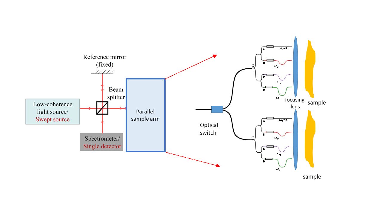
“Taking 100 to 120 reference points, our scanner ‘bundles’ them together, acquiring 20 arms at a time. However, it is our patented switch that moves from bundle to bundle in nanoseconds that gives us the ability to quickly acquire the images in real-time,” said Dr Avci.
Standard OCT works by collecting data from a single sample arm, which is acquired mechanically using a scanner. The final image is formed by combining these individual images during post-processing.
“The OCT we have today uses a process called ‘eye-tracking’ which can be tricky and involves many elements to do it right. However, if we can manage to create an image before the eye moves (in 5-10 sec or so) then there is no need for tracking schemes,” said Dr Avci.
Working with ACTPHAST 4R – an EU innovation hub designed to give researchers working in academia throughout Europe access to top-level expertise and technologies in photonics to produce demonstrators for their scientific breakthroughs, similar to the supports provided by the separate ACTPHAST 4.0 innovation hub for European companies, especially for SMEs – Dr Avci’s team has been able to access the right technical and business coaching expertise to advance the scanner concept towards an actual product.